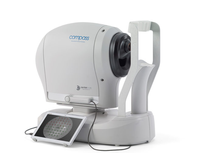
The iCare COMPASS is more than just a Standard Automated Perimeter.
Its built-in innovative retinal tracker gives it a cutting-edge advantage over competition by ensuring a reliable, artifact-free test approach — offering increased clinical value, accuracy, and patient comfort.
Perimetry and fundus imaging are important tools in assisting eye care professionals in detecting and monitoring signs of glaucoma and retinal diseases. However, to ensure optimal accuracy and reliability in results, there is little room for error during examinations – for instance, there is a constant risk of patient eye movements leading to motion artifacts that could affect the results.
Active tracking of eye movements
Retinal tracker is a unique feature at the heart of iCare’s COMPASS automated perimeter which ensures continuous, automated, tracking of eye movements and blinking through infrared retinal scanning. It offers real-time compensation for fixation losses with perimetric stimuli automatically re-positioned before and during projection based on current eye position. This eliminates the risk of motion artifacts.* Stimuli lost due to blinking do not interrupt the test and are automatically repeated during the test.
How does the retinal tracker feature work and what kind comprehensive clinical value does it provide?
iCare COMPASS – the only standard automated perimeter with active retinal tracker
The advantages that iCare COMPASS has over Standard Automated Perimetry include the ability to measure sensitivity at specific retinal locations with high topographic accuracy. This is possible due to retinal tracking which compensates for eye movements during a visual field examination.
Standard automated perimeters in the market cannot actively compensate for eye movements during the test in real time. They either ignore eye movements which could lead to motion artifacts in the visual field results, or they experience data loss when patients blink.
Continuous infrared imaging of the retina
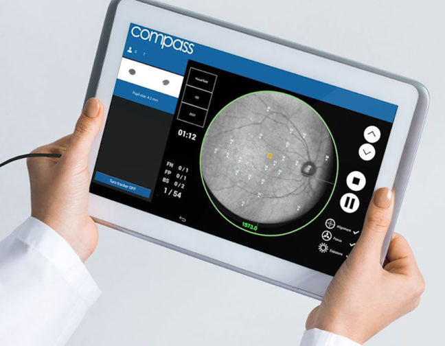
iCare COMPASS images the retina with infrared 25 times a second, making it possible to track even the smallest eye movements and blinking, and compensate for them. The tracker ensures that the next stimulus meant for the same location really is in the same anatomical location, despite eye movements.
The system can be visualized as a retinal map of points, where the stimuli are supposed to appear during the test. When the eye moves, the map of points moves accordingly.
There is no other perimeter in the clinical standard automated perimeter class that has an active retinal tracker capable of compensating for eye movements.
Superior repeatability for effective monitoring of visual field
Apart from combining structure and function, one of the biggest clinical benefits of iCare COMPASS’ retinal tracker is superior repeatability and accuracy of the perimetric test.
As the retinal tracker ensures the test data always comes from the same anatomical locations, iCare COMPASS offers superior visual field test repeatability for effective and reliable long-term monitoring of the retinal defects.
Typically, in retinal defects like glaucoma, it is essential to be able to see pointwise progression as well as a trend of global indices over years. Once baseline tests are recorded, it is important that any further tests with the same test pattern and strategy are without eye movement artifacts and lost stimuli due to blinking. In other words, it is beneficial that the test-retest variability is very low. iCare COMPASS offers this benefit.
Reliable detection and delineation of visual field defects
In patients that have unstable fixation, the retinal tracker offers unparalleled advantage over the competition — reliable detection and delineation of visual field defects.
For instance, it is beneficial to precisely distinguish between absolute scotoma areas and areas with well-preserved vision for reliable follow-up, and for resolving fixation issues in case of central scotomas. It would be impossible to outline these areas precisely without a retinal tracker.
Other standard automated perimeters in the market cannot reliably detect how large or small the defected areas are. The iCare COMPASS’ retinal tracker offers better detection and delineation of these areas to more accurately identify and separate visual field defects.
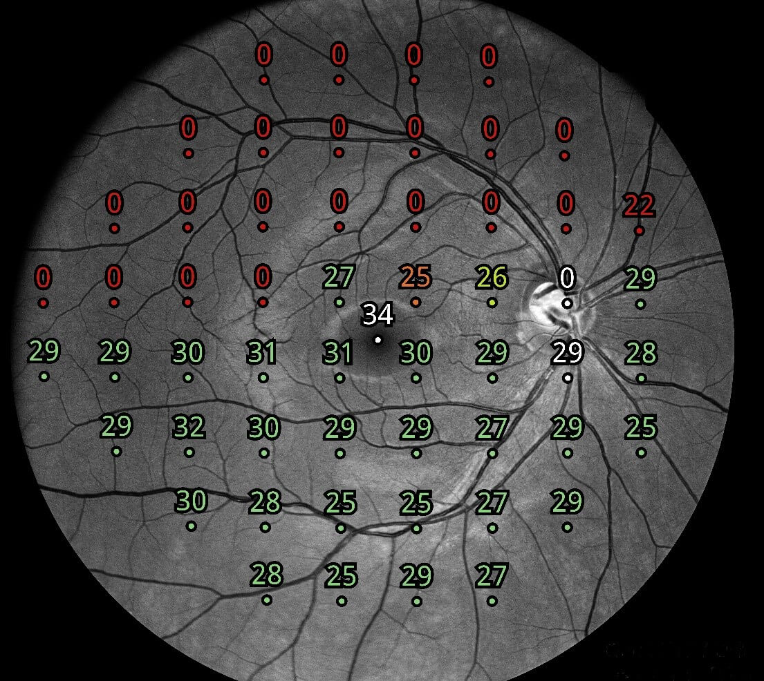
The retinal tracker also offers a better distinction between glaucoma and other diseases, due to its precise structure and function correlation. This is made possible by a combination of three things — visual field test, retinal tracker, and retinal image background, on which the visual field threshold sensitivities are superimposed for better detection of retinal defects and diseases.
Retinal tracker for an improved patient experience
Unlike standard automated perimeters in the market, iCare COMPASS does not have to rely on the objectivity of the operator when there is loss of data or fixation instability. The active retinal tracker ensures test results are always reliable, which benefits the patients by aiding in the diagnostic process more accurately.
The retinal tracker also ensures optimal patient comfort since the test can be carried out quickly without any risk of data loss and without adding to the fatigue of the patient through interruptions every time the patient blinks.
During a COMPASS exam, it doesn’t matter if the patient blinks
During a COMPASS exam, it doesn’t matter if the patient blinks. It is the only standard automated perimeter that goes on and repeats the lost stimuli without the patient even knowing about it, because of the retinal tracker. Patients can also rest by closing the eye and pulling away and continue at any time without loss of data.
iCare COMPASS is the first standard automated perimeter that performs visual field tests using a real-time retinal tracker while delivering ultra-high-resolution confocal TrueColor fundus images at the same time. This increases clinical value, patient comfort and correlation between retinal sensitivity values and retinal structure.